Home
/ Structure Plant Cell Under Microscope / How To See A Plant Cell Under A Compound Microscope Quora - Start studying cell structure & microscopes.
Structure Plant Cell Under Microscope / How To See A Plant Cell Under A Compound Microscope Quora - Start studying cell structure & microscopes.
Structure Plant Cell Under Microscope / How To See A Plant Cell Under A Compound Microscope Quora - Start studying cell structure & microscopes.. It also has a very high resolving power. Within the cytoplasm, the following organelles are visible in almost all cells except prokaryotes when looking at higher magnification (ie endoplasmic reticulum studded with ribosomes looks rough under the microscope; We say cells are microscopic because they can only be seen under a microscope. They lack any internal structure and contain hydrolytic enzymes. Their distinctive features include primary cell walls containing cellulose, hemicelluloses and pectin, the presence of plastids with the capability to perform photosynthesis and store starch.
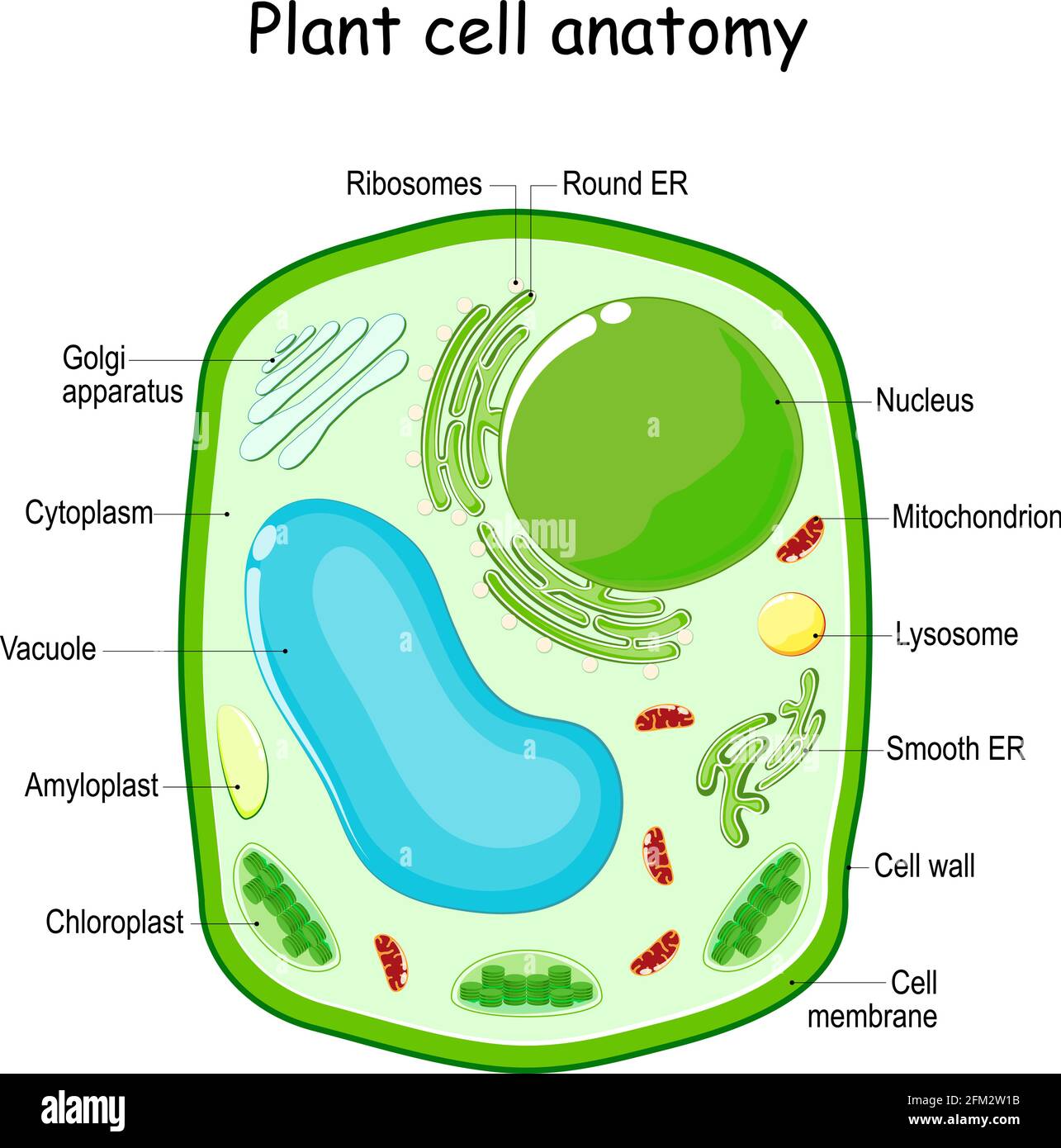
All living things are made up of one or more cells the cell is the. Identify two functions of plastids in plant cells. Plant cells have cell walls, one large vacuole per cell, and chloroplasts, while animal cells will have a cell membrane only. Plant cells also usually have a distinct shape. Here's a diagram of a plant cell:
Plant Cell High Resolution Stock Photography And Images Alamy from c8.alamy.com Structure and functions of the cell organelles. Plant cells also usually have a distinct shape. They can be observed under electron microscope only. Plant cells have a regular shape and structure and keep their shape easily. Examining plant cells under the microscope. Body cells under a microscope. Dreamstime is the world`s largest stock photography community. Return from epidermal cells to microscopemaster home.
Use them in commercial designs under lifetime, perpetual & worldwide rights.
The rigid exterior around the cells is necessary to allow the plants to list three structures that are found in plant cells but not in animal cells. Your plant cells under microscope stock images are ready. There are various tasks done by a cell to complete them as the cell is the basic purposeful and. The plant cell as more rigid and stiff walls. The type of cell and the structure of cells. These are both specific types of cells, and. Learn vocabulary, terms and more with flashcards structure: Their distinctive features include primary cell walls containing cellulose, hemicelluloses and pectin, the presence of plastids with the capability to perform photosynthesis and store starch. Robert hooke was the first cytologist to identify cells under his microscope in 1665. Start studying cell structure & microscopes. Plant cells have a regular shape and structure and keep their shape easily. It also has a very high resolving power. Identify two functions of plastids in plant cells.
Pupil activity • cell structure • read through the information on each of the organelles. They are the main sites of hydrolytic enzymes and so can. It's amazing how much is hidden from the human eye and exists at the level of the microcosm! The differences between plant and animal cells. Green plant cells under microscope.
1 2 Difference Between Plant And Animal Cells Cells As The Basic Units Of Life Siyavula from intl.siyavula.com Resolving power is the ability to distinguish between separate things which through the electron microscope, very fine details of the cell can be observed. Here's a photo of a plant cell under an electron microscope. Cell structure view of leaf surface showing plant cells under microscope for education. Examining plant cells under the microscope. To examine a specimen like plant or animal cells under a microscope you need to prepare microscope slide. Download this microscopic image of a buttercup plant photo now. What are the roles of the. Published on december 9, 2013 at 4:03pm by glenda stovall under cell.
It also has a very high resolving power.
Body cells under a microscope. The plant cell as more rigid and stiff walls. To study the microscopic structures of human cheek cells under a compound microscope. A micrograph is a photo or digital image taken through a microscope to show a magnified image of a specimen. Examining plant cells under the microscope. Download this microscopic image of a buttercup plant photo now. 8 pictures of plant cells under a microscope. The features are the structures of bacteria and fungi. Green plant cells under microscope. It also has a very high resolving power. There are various tasks done by a cell to complete them as the cell is the basic purposeful and. Plant cells also usually have a distinct shape. (a) state the resolution and magnification that can be achieved by a light microscope, a transmission electron microscope and a scanning electron (j) compare and contrast, with the aid of diagrams and electron micrographs, the structure and ultrastructure of plant cells and animal cells.
The plant cell as more rigid and stiff walls. Many plant cells are green. 8 pictures of plant cells under a microscope. A cell is a very tiny structure which exists in living bodies. Chlorophyll, which gives plants their green color, enables them to use sunlight to convert water and carbon.
Plant Cell Structure Cross Section With Green Cell Wall Membranes And Chloroplasts Purple Nucleus Stock Vector Vector And Low Budget Royalty Free Image Pic Esy 042371786 Agefotostock from previews.agefotostock.com This gives rise to its name of rough endoplasmic reticulum. Robert hooke was the first cytologist to identify cells under his microscope in 1665. Plant cells have cell walls, one large vacuole per cell, and chloroplasts, while animal cells will have a cell membrane only. A microscope is an important instrument for studying cells e.g. Structure and functions of the cell organelles. (a) state the resolution and magnification that can be achieved by a light microscope, a transmission electron microscope and a scanning electron (j) compare and contrast, with the aid of diagrams and electron micrographs, the structure and ultrastructure of plant cells and animal cells. Return from epidermal cells to microscopemaster home. There are various tasks done by a cell to complete them as the cell is the basic purposeful and.
This means a cell does various tasks that it was designed to accomplish.
Return from epidermal cells to microscopemaster home. To examine plant cells under a microscope and find and identify different cell parts. The type of cell and the structure of cells. What are the roles of the. They can be observed under electron microscope only. Cells ultrastructure of a plant cell as seen through an electron microscope. Dreamstime is the world`s largest stock photography community. Published on december 9, 2013 at 4:03pm by glenda stovall under cell. Many plant cells are green. Plant cells are eukaryotic cells with a true nucleus along with specialized structures called organelles that carry out certain specific functions. some of these differences can be clearly understood when the cells are examined under an electron microscope. For example, the main parts of the plant cells different from those in animals. The plant cell as more rigid and stiff walls. Although they serve a number of important functions, their primary role is to protect from a variety of harmful return to leaf structure under the microscope.
Share :
Post a Comment
for "Structure Plant Cell Under Microscope / How To See A Plant Cell Under A Compound Microscope Quora - Start studying cell structure & microscopes."


Post a Comment for "Structure Plant Cell Under Microscope / How To See A Plant Cell Under A Compound Microscope Quora - Start studying cell structure & microscopes."